Biomaker

sciTAG: a label design and printing app created by scientists for scientists

Proper labelling practices in the lab is one of the first things students are taught at the practical courses. As a matter of fact, labelling is an essential part of data management for scientists while working in the laboratory. And if you ever worked in a lab, you must know how much time you usually spent to label every single tube, falcon, vial, bottle etc.

The Problem

Appropriate labelling is an essential research practice. It is fundamental to day-to-day operation and long-term sample storage in laboratories. Creating labels with handwriting is convenient, adaptable, and often the standard method scientists use. However, variabilities in label format and illegible messy handwriting can both lead to flawed and/or erroneous communication, resulting in, for example, loss of important biological samples. Moreover, writing labels by hand for large scale experiments or a high number of samples is time-consuming and creates ergonomic stress. The problem presented called for a labelling method that is automated, user-friendly, and affordable.

The Project

We proposed to develop an IOS app that allows biological and medical laboratories to operate in a consistent and efficient manner, by making label design and printing more accessible.

We envisaged an app which is user-friendly, automated, and compatible with affordable label printers. To accomplish our goal, we followed a stepwise approach composed of three parts: user interface design, technical structure of the app and testing the app.

Efficient label creation frees scientists from the laborious task of writing labels by hand. Consistently and systematically designed labels ensure samples stored for long term are readable and used accurately. The app will be free, meaning that convenient label printing will be accessible to the wider scientific community.

What Has Been Achieved So Far?

Our team of five is composed of a molecular biologist, a microbiologist, a geneticist, a bioinformatician and a software developer. We started by self-teaching ourselves how to design and develop an app since most of us never worked on app development before.

Interface design decisions were made not only to be attractive to potential users, but also to be functional and simplistic as possible. One aspect of the design is visual elements. We decided on aesthetic choices such as a colour scheme throughout the app. We considered colour blindness since 4.5% of the global population experience colour-blindness. Next, we decided on the name of the app: sciTAG. We did market research to make sure this name is not a trademark. Finally, we designed an app logo depicting a double-stranded DNA alongside the name of the app.

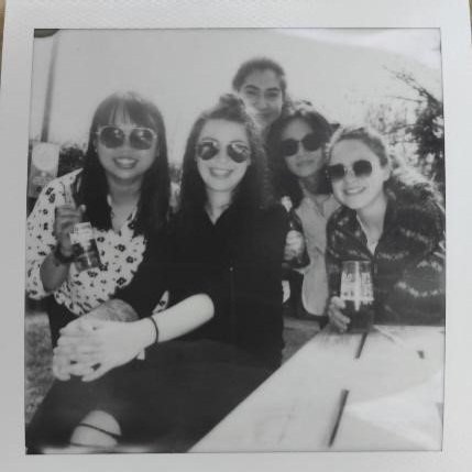
From left: Junyan Liu (Postdoctoral Researcher, Sanger Institute), Marta Matuszewska (PhD student, Department of Veterinary Medicine, University of Cambridge) Begum Akman (Research Associate, Department of Pharmacology, University of Cambridge), Chuqiao Gong (Software developer, EMBL-EBI), Ellis Kelly (PhD student, Department of Genetics, University of Cambridge)

Then, we identified the features that are crucial for our target audience. We divided the label design into two blocks by keeping it as simple as possible with choices included to create a good label.

From left to right: home screen, label design screen, prototype label in PDF format.

Next step was to layer the technical structure: first user input information used to create a printable object such as PDF. Second, to convert this object to a storable object within the app. Finally, the app had to be compatible with commercially available label printers.

What’s Next For The sciTAG Team?

With the help of the Biomarker Challenge funding, we sourced several label printers available in the market to test sciTAG app. Among these printers only one of them is specific for lab settings (very expensive!) and others are highly affordable printers that we sourced from different companies. We distributed these printers within the team, and we are planning to complete the sciTAG prototype, start testing and reporting our experiences.

After the successful tests from the team, we are planning to recruit five laboratories within University of Cambridge to further test and optimize the sciTAG app. Eventually we are aiming to make sciTAG freely available to the science community on the App Store. Further down the line we also would like to consider making our app available for android users.

Developing an open and affordable 3D bioprinter

Background

Friends had Central Perk cafe, the gang from How I Met Your Mother had MacLaren’s Pub and we have Charlie’s Pizza joint. What started as a trivial discussion about printing human organs over a stone-baked Margherita quickly evolved into us applying for a bid to participate in the BioMaker Challenge, with the objective of Developing an open & affordable 3D bioprinter

The beginnings of 3D printing date back to the 1980’s, when it was commonly known as its more mouthful synonym stereolithography (SLA), a technology pioneered by Charles W. Hull. Revolutionized with SLA, one could translate a 3D design from a data file into a physical object, in a relatively short time. The household notion of 3D printing is testament to the success of SLA, which originated from the company 3D Systems Corporation. 

Fast forward 40 years, my neighbours are at home 3D printing little elves for their garden, my cousin is buying a 2-story building printed by an enormous SLA machine, my doctor is offering his patients custom 3D printed ear implants, heart valves or bones - SLA has already infiltrated many aspects of our lives, economy and medical care. Much of this progress came about because of the RepRap project, whose aim was to create an open-source 3D printer capable of printing most of the parts needed to replicate itself, making it cheap enough for hobbyists. Today’s consumer 3D printer companies, and the widespread use of 3D printing, grew out of the RepRap hobbyist community. 

The field of regenerative medicine could hugely benefit from 3D printing techniques to offer personalized medicine to its patients, by for example printing organs made of one’s own cells. However, these applications are challenged by the complex architecture of human organs, the difficulties of supporting living cells, introducing foreign materials in a human body and working at a microscale. 

Addressing these issues, companies and academic researchers have built 3D printing platforms, specialized for biological materials, hence 3D bioprinting. Not surprisingly, these come at a prohibitive cost, up to £200K for the printer only.

BioMaker Challenge

During our weekly pizza gatherings we talked about how it would be great to have an open-source bioprinting project, along the same lines as the original RepRap project. It could make the technology more accessible and bring printing human organs for transplantation a step closer. None of our backgrounds were in bioprinting, but our combined skillsets did range from engineering and programming to cell biology, with diverse backgrounds in start-ups, industry and academia. The Biomaker challenge would be a great way to work together on a project where we could contribute something to the 3D printing community, and learn a lot along the way. Plus it would be fun! 

3D bioprinter.

Bertie the 3D bioprinter.

So as part of the BioMaker Challenge, we set out to address the lack of reasonably priced open-source 3D bioprinters in the market by Developing an open & affordable 3D bioprinter. With this project still in its infancy, working with human cells was out of the scope due to ethical and practical reasons. Nevertheless, we attempted to design our 3D printer to potentially print corals, an application with a shorter timeline, more amenable to working in the absence of a biological laboratory and with direct benefits to a problem close to our hearts, the depreciation of the ocean’s corals. Our project branched into two components outlined below: 

1. Converting an existing open-source 3D printer into a 3D bioprinter capable of extruding biomaterial.

Our inspiration to address this first problem is drawn from a paper by an American group (Push et., 2018), in which they describe the design of a syringe pump large volume extruder (LVE) of low cost, compatible with printing biomaterial. This involves the purchase of standard materials (e.g: 60mL syringe, bolt nuts) and the 3D printing of other assembly components with a PLA extruder from a standard 3D printer. The newly assembled LVE would then replace the printer’s standard PLA extruder and allow the printing of biomaterials. 

With this in mind, many hours of research and discussions went into selecting the right 3D printer as a foundation for our bioprinter - we were certain about two things: it had to be open-sourced and stay within a reasonable budget. We finally settled on the RepRap Ender 5 3D-printer. In addition to the syringe extruder, we were interested in trying to integrate a peristaltic pump in our system because it would allow us to extrude multiple bio-inks at once. 

Adding a valving architecture between the reservoirs and the pump enables the switching of the reservoir that bioinks are aspirated from on the go, thereby enabling the interlinking of multiple inks of different cell types in 3D space. 

After a few months of putting all the hardware together and with a sprinkle of software, Bertie the Bioprinter was born!

2. Developing a bioink suitable for sustaining living cells.

Regardless of the organism in question, a bioink should: 

a) Contain the right nutritional environment for specialized cells.

b) Maintain a single-cell suspension (prevent cells from clumping together and/or from dying from being separated from their friends).

c) Provide a physical scaffold once printed to keep the shape of the 3D model.

d) Use materials that are safe to insert in the receiving environment. 

Printing human cells was out of the scope of Biomaker, so we initially turned our attention to another challenging bioprinting application, trying to 3D print coral (which not all of us knew was a living organism!). Coral numbers and health have declined rapidly as a result of environmental conditions and their slow growth cycle. If coral could be 3D bioprinted, perhaps it could be transplanted back into reefs to bring them back to life.

We dove into the scientific literature and identified two such bioink formulas, which are, in theory, capable of supporting the life cycle of corals and providing an “ocean-friendly” physical scaffold. It turned out however that coral species are high maintenance, requiring expensive aquariums and daily care. Even the most experienced coral growers find it challenging to maintain a community of the families of coral that were the right size to fit our bioprinter. We realised that we didn’t have the funds or the time to manage coral during the Biomaker Project, but we did make connections with coral researchers who we hope to work with on this project in the longer term. 

Bertha.jpg

Printed phytoplankton using Bertha the bioprinter.

We were however determined to evaluate the efficacy of our bioinks, and didn’t let coral’s needy demands deter us. One of the bioinks we identified is based on the chemical interaction of sodium alginate and calcium chloride, which create a solidified gel upon contact. At the concentration used, these chemicals didn’t affect the survival of phytoplankton, a single cell organism we chose to work with for its relative low cost, ease of maintenance and ability to survive as single cells. 

By the end of the project we were able to print a very slimy collection of phytoplankton, and affectionately named it Bertha. Bertha still sits in someone’s fridge for posterity.

Future

The BioMaker Challenge was an incredible opportunity for us to concretise an idea and start along the path to our long term goal. We also got to meet a wonderful community of makers and with their help and feedback we hope to further refine Bertie the Bioprinter. Moving forward, we plan to improve Bertie’s performance in terms of printing precision and its ability to run complex modes (e.g: multiple bioinks printing at once), as well as make him suitable for sterile work (e.g: ventilation, UV lights). We also plan to develop  application specific bioinks for Bertie to print. 

We’d like to take this opportunity to thank the BioMaker Organisers (special thanks to Alexandra, Jenny & Jim) for arranging this event, Professor Ludovic Vallier (Cambridge Stem Cell Institute, Jeffrey Cheah Biomedical Centre) for supporting our ideas and administrative needs as well as the BioMakeSpace Cambridge who welcomed us in their community.  Thanks to the BioMaker Challenge, we are now one small step closer to 3D printing organs, and in the meantime, Bertie is on standby for printing COVID-19 related materials (with the original PLA extruder) if need be. 

Julie, Sebastian, Monica, Robin, Ben & Tejas

PS, For more details of the project, check out our Hackster :-)


OpenCM - an open framework for single cell manipulation

Introduction

Microscopic studies of single cells, their dynamics, signalling and behaviour require a whole toolbox of different microscopic techniques, highly specific gadgets and methods that can easily exceed the budget for new lab equipment.

OpenCM team.jpg

OpenCM team (left to right): Maziyar Jalaal, Nico Schramma, Stephanie Höhn and Kyriacos Leptos

The open technology community is continually developing new, innovative and inexpensive solutions for easy-to-build microscopes and other equipment, aiming to democratize microscopy and enhancing the applicability of custom-built devices to approach novel questions. The OpenCM team aimed to provide one of the solutions needed to make open-source microscopes even more utile, namely a setup for micromanipulation.

Multi-axis micromanipulation is used for in-vitro fertilization, imaging of highly motile cells, microinjections, patch-clamp methods, mechanical probing of cells with micropipettes and is - simply put - the microscopists tiny helping hand to interact with the sample mechanically.

The project

Key to the OpenCM project is the close collaboration with Benedict Diederich and René Lachmann from the UC2 group, which started after the OpenPlant forum in July 2019. UC2 builds 3D-printed modular optical elements, which can be easily assembled into various microscopes. The OpenCM project aims to make this system even more versatile, by adding a module for micromanipulation.

During the BioMaker challenge the team was able to design the first prototype two-axis micromanipulator, which fitted the UC2 system and was controlled via Arduino. The team followed the online documentation of their collaborators in order to 3D-print a microscope, which is controlled with an open source Python based GUI on a Raspberry Pi. The team was among the first to reproduce the UC2 microscope and helped evaluate and enhance their current quality of the online documentation.

In the end, the team was able to build a fully integrated microscope and micromanipulation system and to showcase it at the BioMaker Fayre. The most important outcome of the BioMaker Challenge is establishing a very close collaboration with the UC2 project, which is still persisting.

Future work

The team plans to undertake the final steps in developing a 3-axis micromanipulator (OpenCM2). The hardware will be integrated into the UC2 system, and free software will be provided for position controlling with a wireless joystick and the UC2 GUI via Raspberry PI and wifi controllers.  

First prototype as presented at the BioMaker Fayre 2019, an inverted microscope with 2 axis micro manipulator.

First prototype as presented at the BioMaker Fayre 2019, an inverted microscope with 2 axis micro
manipulator.

The project will continue in close collaboration with the laboratory of UC2 in Jena, Germany, and the financial support in the context of the programme “Creativity and Studies” of the University of Gottingen by the AKB Stiftung, a non-profit foundation of the Büchting Family (secured by Nico Schramma and Björn Kscheschinski).

Furthermore, the team found the UC2 setup very useful to provide hands-on experience at the intersection between physics, mechanical engineering, microscopy and cell biology. Hence, as a side project, the team also started preparing workshops for children and students.

In a joint effort, and with the financial help of the UK's Women Engineering Society and the Public Engagement office at the University of Cambridge, the members of UC2 and OpenCM participate at the Women in Engineering Society 100 Violets Challenge in order to give a first "Optics for Everybody" workshop. Unfortunately, due to the pandemic, the exhibition at the Brunel Museum in London has been postponed.

During the BioMaker challenge and driven by the community's open-mindedness, the team was encouraged to branch out ideas, communicate openly, and team up with others to find and combine new and innovative solutions. In the end, the BioMaker spirit inspired the OpenCM team to reach out to the broader community.

Acknowledgments

We thank David Page-Croft for fruitful discussions and his help with 3D printing. We also thank Benedict Diederich, René Lachmann and Barbora Marsikova from the UC2-Project, and Alexandra Ting, Dieuwertje van Esse - van der Does and Jim Haseloff from the BioMaker Challenge.

Written by Maziyar Jalaal, Nico Schramma, Stephanie Höhn and Kyriacos Leptos.

References

Diederich, Benedict, et al. "UC2-A Versatile and Customizable low-cost 3D-printed Optical Open-Standard for microscopic imaging." bioRxiv (2020).


Digital Workshop: No-Code Programming for Biology

Sadly, in order to protect our community, the popular No-Code Programming for Biology workshop due to be held on 23rd-24th March was cancelled. However, we’re pleased to announce that the materials for this workshop are now being made available online.

no-code_logo2.png

This course was designed to introduce biologists to the basics of building custom instrumentation to assist with experiments in the lab and field. Part of the long-running Biomaker project, the course covers the fundamentals of using Arduino-based microcontrollers, sensor electronics, displays and actuators, as well as use of the visual programming language XOD. By combining these skills participants can learn to build a variety of instruments that are useful for measuring and controlling biological systems.

Making your own devices for use in biological research can be advantageous in many ways – components are often very cheap compared to commercially available options; there is a well-established community of experienced scientists focused on collaboration and open sharing of designs; and the opportunities for customisation and experiment-specific adjustments are endless. However, there are also hurdles for those looking to build custom instruments. Without a background in electronics or engineering it can be hard to know where to start. In addition, many systems require the maker to be able to understand and write code in languages such as CC+, which take time to learn and contain complex syntax.

Biomaker and the No-Code Programming for Biology workshop aim to break down some of these barriers. The course provides biologists with a hardware starter kit, a series of simple tutorials to get started using hardware and electronics, and an introduction to the visual programming language XOD. Instead of having to write code to control your instruments, XOD uses ‘nodes’ to represent different hardware and functions. Connecting these nodes in different combinations allows you to control your hardware and customise your instrumentation.

Previously, teams working on Biomaker projects have used these concepts for a wide variety of applications, including instrumentationmicroscopymicrofluidics3D printingbiomedical devicesDNA designplant sciences and outreach and public engagement. The No-Code programming for Biology workshop builds on what we have learned from running Biomaker projects and provides biologists with the necessary skills to start building their own devices and advance their research with inexpensive custom instrumentation.

If you would like to get involved with the No-Code Programming for Biology course the introduction, and first set of tutorials are now available on the Biomaker website. A number of hardware starter kits may be available upon request, although this cannot be guaranteed.

More information about the Biomaker project can be found on the Biomaker website. For questions and enquires please contact the Cambridge SynBio IRC and OpenPlant Coordinator Steph Norwood at coordinator@synbio.cam.ac.uk.